Case Study: Femur Analysis with UKE, Hamburg
In collaboration with the Institute of Osteology and Biomechanics, UKE we used XamFlow and CLIP to analyse subchondral cysts in femoral heads.
“I’m really impressed!” – Felix Schmidt, Institute of Osteology and Biomechanics, UKE
Dataset
Multiple samples of femoral head measured with SCANCO XtremeCT II
17 GB per image
6 images analysed initially
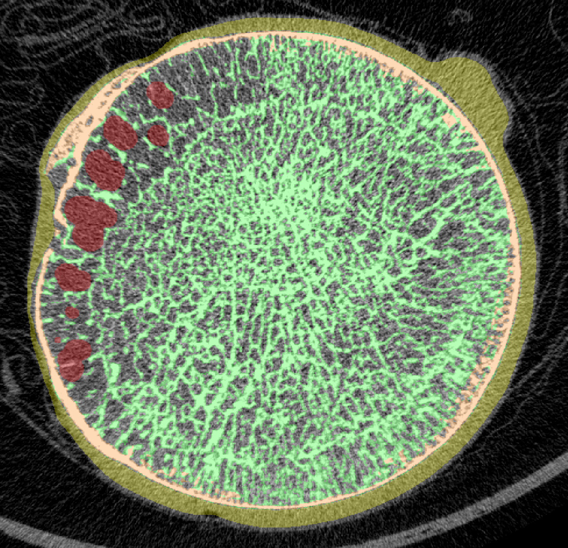
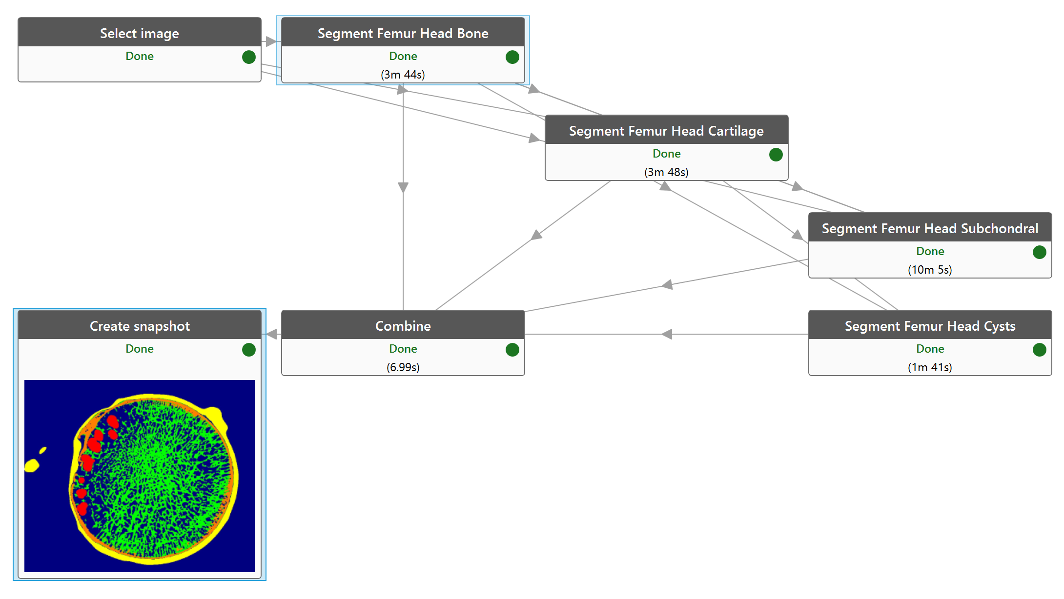
Workflow
We used CLIP to implement four stages in a multi-object segmentation workflow:
Bone mask and hull
Cartilage mask and loss area
Subchondral area mask
Cysts
The CLIP curvelet filter was used to identify the subchondral area.
References
Rossinelli, D. (2019). CLIP
Chiba, K., Burghardt, A. J., Osaki, M., & Majumdar, S. (2014). Three-dimensional analysis of subchondral cysts in hip osteoarthritis: an ex vivo HR-pQCT study. Bone, 66, 140–145. https://doi.org/10.1016/j.bone.2014.06.001